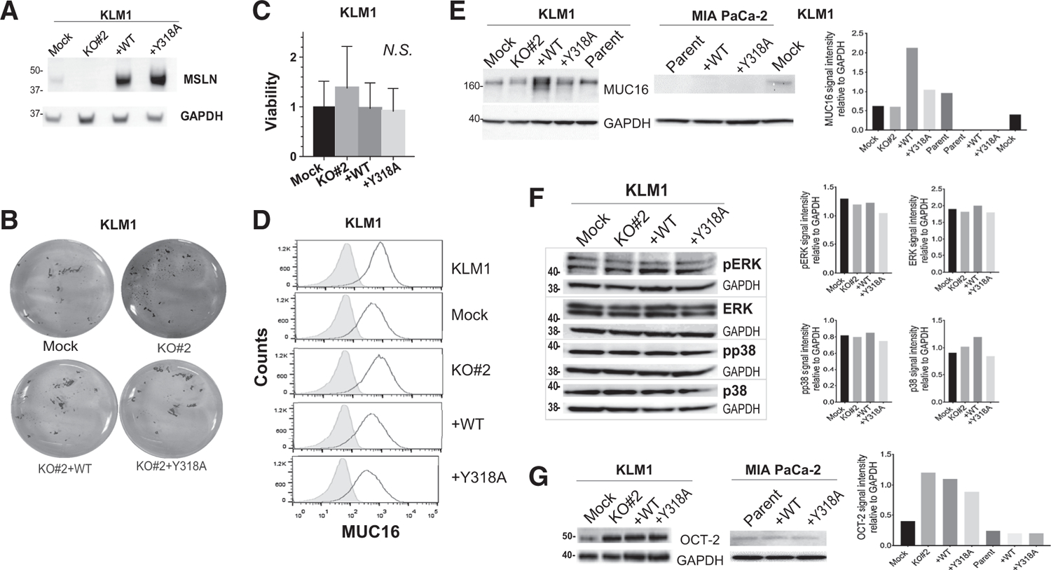
Figure 5.

Identifying a mechanism for MSLN tumorigenicity. A, Lysates from harvested KLM1 intraperitoneal tumors were immunoblotted with anti-MSLN antibody to demonstrate continued expression of the transgene in vivo. B and C, Tumor cells were plated at equal numbers onto ultra-low adherence plates. B, After 24 hours, the cells were stained with crystal violet and photographed. C, Viability was measured by colorimetric assay after 72 hours. N.S. = no significant difference. D, Cell membrane expression of MUC16 was measured by flow cytometry (black outline). Shaded peak shows control where primary antibody was omitted. Immunoblots were performed to examine: E, Total MUC16 expression and associated quantitation (F) total and phosphorylated ERK and p38, and (G) total OCT-2 expression and associated quantitation in KLM1 and/or MIA PaCa-2 cultured cell lysates. GAPDH was used as loading control in A and E–G.
