Figure 4.

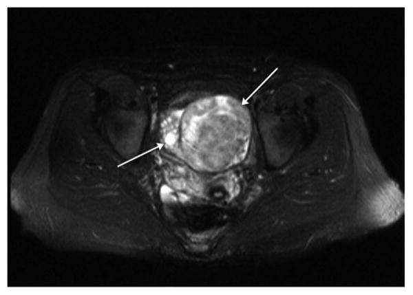
T2 weighted axial MRI scan shows heterogeneous mass replacing left ovary (long arrow) that was a primary ovarian lymphoma. Adjacent normal right ovary is seen (short arrow).

T2 weighted axial MRI scan shows heterogeneous mass replacing left ovary (long arrow) that was a primary ovarian lymphoma. Adjacent normal right ovary is seen (short arrow).