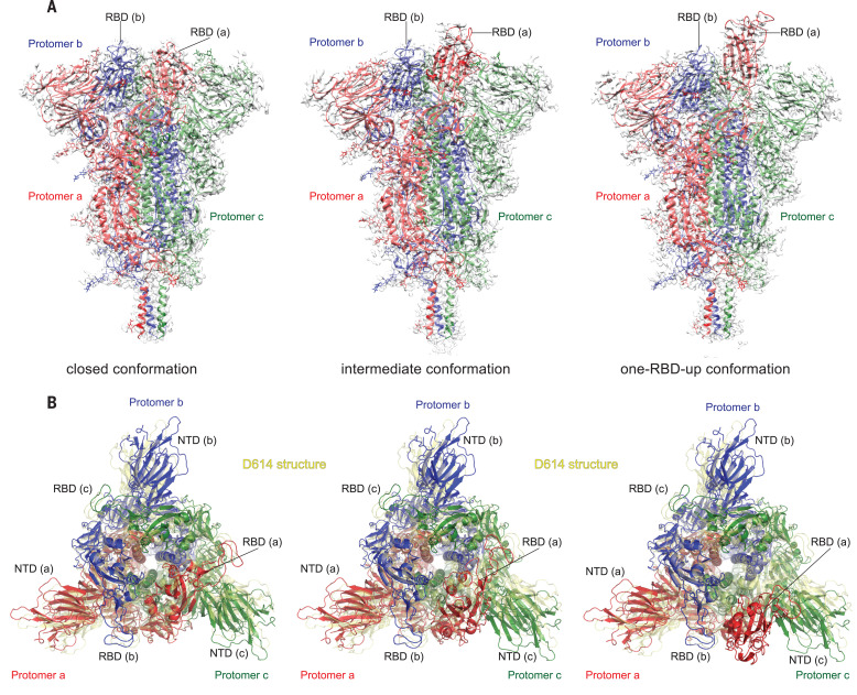
Fig. 2. Cryo-EM structures of the full-length SARS-CoV-2 S protein carrying G614.
(A) Three structures of the G614 S trimerrepresenting a closed, three RBDdown conformation; an RBD-intermediate conformation; and a one RBDup conformationwere modeled on the basis of corresponding cryo-EM density maps at 3.1- to 3.5- resolution. Three protomers (a, b, and c) are colored in red, blue, and green, respectively. RBD locations are indicated. (B) Top views of the superposition of the three structures of the G614 S in (A) in ribbon representation, with the structure of the prefusion trimer of the D614 S (Protein Data Bank ID: 6XR8) shown in yellow. The NTD and RBD of each protomer are indicated. Side views of the superposition are shown in fig. S8.

