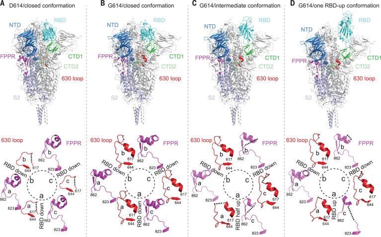
Fig. 3. Cryo-EM structures of the full-length SARS-CoV-2 S protein carrying G614.
(A) (Top) The structure of the closed, three RBDdown conformation of the D614 S trimer is shown in ribbon diagram with one protomer colored as NTD in blue, RBD in cyan, CTD1 in green, CTD2 in light green, S2 in light blue, the 630 loop in red, and the FPPR in magenta. (Bottom) Structures of three segments (residues 617 to 644) containing the 630 loop in red and three segments (residues 823 to 862) containing the FPPR in magenta from all three protomers (a, b, and c) are shown. The position of each RBD is indicated. (B to D) Structures of the G614 trimer in the closed, three RBDdown conformation, the RBD-intermediate conformation, and the one RBDup conformation, respectively, are shown, as in (A). Dashed lines indicate gaps.

