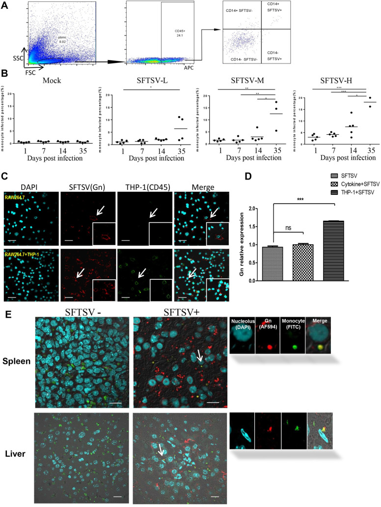
Fig 3. HuPBL-NCG mice increase SFTSV susceptibility in host tissues.
(A) Flow cytometry analysis of the percentage of CD14+ human monocytes among SFTSV infected cells. (B) At different time point, the percentage of virus-infected monocytes among infected with different virus titer in HuPBL-NCG mice. (C) THP-1 significantly improve the virus infection ability among mouse RAW264.7 cells in vitro. Bar = 50 μm. (D) Relative expression levels of viral mRNA in RAW264.7 cells in different co-cultured assays. (E) SFTSV infected cells in HuPBL-NCG mice detected by IFC on sacrifice day. Representative tissue sections of spleen and liver were analyzed by antibodies specific to SFTSV Gn in red and to human CD14 in green among three groups of mice. Nucleoli was stained by DAPI in cyan. Bar = 20 μm. The enlarged images of individual cells were pointed by corresponding arrows. The images are 600 x. Data are shown as mean±SEM of three independent experiments. (*p <0.05, **p <0.01, ***p <0.005).

