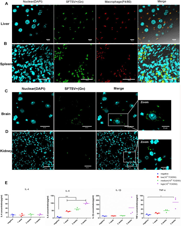
Fig 4. Virus localization and proinflammatory cytokine determination in infected animals.
(A) SFTSV and macrophages were identified by immunofluorescence in the liver of SFTSV-infected mice. Experiments were performed with three independent trials and similar results were reproduced. Scale bars, 10 μm. (B). Confocal microscopy to examine colocalization of SFTSV (in green), and macrophages (in red) in the SFTSV-infected spleen. Scale bars, 30 μm. (C and D). The presence of the SFTSV Gn antigen was confirmed in the brain and kidney. Scale bars, 20 μm and 30 μm, respectively. (E). Kinetics of proinflammatory cytokines in SFTS virus–infected mice. Plasma levels of cytokines were quantified by ELISA assays between the infected group (105 TCID50) and the control group (negative) after an indicated time. IL-1β, interleukin 1β; IL-4, interleukin 4; IL-6, interleukin 6; TNF-α, tumor necrosis factor α. Data are shown as mean±SEM of three independent experiments. (*p <0.05, ***p <0.005).

