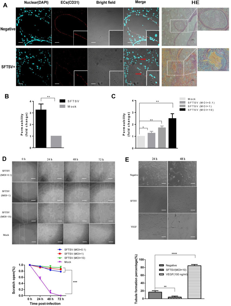
Fig 5. SFTSV infection disrupts the endothelial cell barrier.
(A) Immunofluorescence staining and H&E staining lung tissues of mice infected with SFTSV on sacrifice day. The continuity and integrity of endothelial cells markedly decreased compared with negative control mice along with a higher incidence of red blood cell leakage around the disintegrated pulmonary vessel as shown by the red arrow. The fluorescent images and H&E images are 400× and 200×, Bar = 50 μm and 100 μm respectively. (B) The extravasation of Evans Blue dye from vessels in the lungs of a mouse can be quantified by Miles assay in vivo. Virus infection has a promotive effect on microvascular permeability with 3.2-fold increased leakage occurred in lung tissues compared with lung injected with saline only. (C) In vitro permeability was measured in HUVEC monolayer cells stimulated with different titers virus. (D) Scratch wound assay infected with SFTSV MOI = 0, 0.1, 1, or 10, respectively, was showed with time-points indicated. Original magnification was 40 x and quantifications of wound closure were shown in the graph below, bar = 125 μm. (E) Tubule formation of HUVECs were analyzed with the presence of SFTSV or VEGF. The results were quantified by counting the tube formation branch point as shown in the beneath graph, scale bars = 50 μm. Data are shown as mean±SEM of three independent experiments. (*p <0.05, **p <0.01, ***p <0.005, ****p <0.001).

