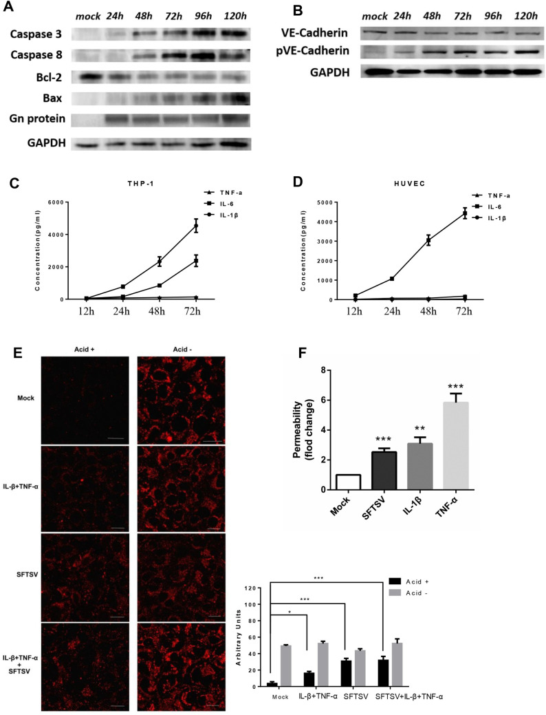
Fig 6. Apoptosis and VE-cadherin internalization were enhanced in pathogenic SFTSV-infected endothelial cells.
(A) High-dose SFTSV replication in HUVECs inducing apoptosis started from 48 hpi. (B) Phosphorylation of VE-cadherin Tyr685 was induced by SFTSV, and contributes to the proper induction of vascular permeability in vitro. (C and D) 3 proinflammatory cytokines released from SFTS virus–infected cells. Supernatant levels of cytokines were quantified by ELISA assays. IL-1β, interleukin 1β; IL-6, interleukin 6; TNF-α, tumor necrosis factor α. (E) Immunofluorescent staining of HUVECs infected with SFTSV or SFTSV-inducible inflammatory cytokines. Following acid washing, the red fluorescent region indicates VE-cadherin endocytosed at higher rates in the virus infected HUVECs. The results were quantified by counting average fluorescence intensity using Image J software. The images are 400×. (F) Confluent HUVECs were infected in triplicate with pathogenic SFTSV at an MOI of 10 or proinflammatory cytokines or mock infected. Either SFTSV alone or SFTSV-inducible inflammatory cytokines could induce endothelial hyperpermeability in vitro. Data are shown as mean±SEM of three independent experiments. (*p <0.05, **p <0.01, ***p <0.005).

