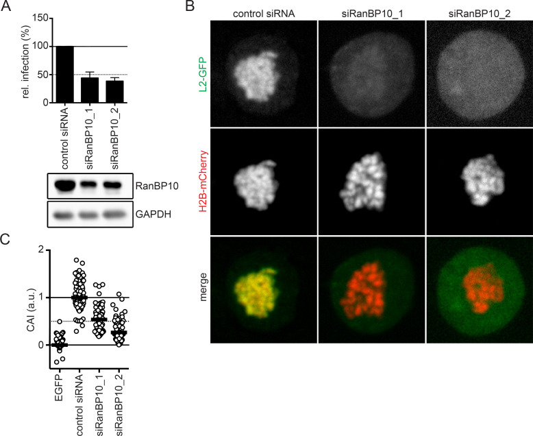
Fig 2. A crucial role of RanBP10 for HPV16 infectivity and L2 tethering.
(A) RNAi of RanBP10 in HeLa cells was followed by HPV16-PsV infection for 48 hours. Infectivity was scored by flow cytometry based on the percentage of the cells expressing GFP. The infectivity was normalized to control siRNA transfected cells and depicted as relative (rel.) infection. The protein expression level of RanP10 upon siRNA knockdown was analyzed by Western blotting. (B) RNAi of RanBP10 in HeLa Kyoto_H2B-mCherry_L2-GFP cells was followed by arrest in prometaphase using nocodazole (330 nM) for 16 hours. Cells were fixed and images were acquired using a spinning disk confocal microscope. Depicted are single median slices. (C) Analysis of (B) quantifying the degree of chromosomal association as described in material and methods. Displayed is the chromosomal association index (CAI) relative to control siRNA-treated cells (1) and GFP expressing cells (0). In three independent experiments, at least 50 cells were analyzed. The median was indicated by a black bar.

