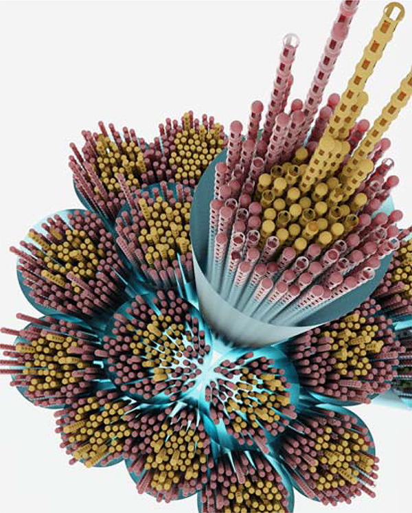
Figure 8.
Illustration showing the fascicular organization of a large diameter zonular fiber. Large fibers are formed from bundles of smaller diameter fibers, each of which contains many individual microfibrils. Bundles are shown surrounded by a glycan coat (blue, see section 2.1) and with a core of fibrillin-2-rich microfibrils (yellow) surrounded by a cortical layer of fibrillin-1-rich fibrils (red). Note that it has yet to be confirmed that the coaxial organization of fibrillins found in the mouse zonule is a universal feature.

