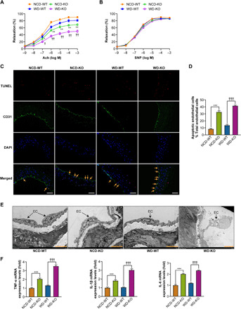
Fig. 1. Myeloid cell–specific MYDGF deficiency is associated with endothelial injury and inflammation in mice.

KO and WT mice aged 4 to 6 weeks were divided into four groups (NCD-WT, NCD-KO, WD-WT, and WD-KO mice) and were fed their respective diets for 12 weeks (10 mice in each group). (A and B) The vasodilation responses to (A) acetylcholine (Ach) and (B) sodium nitroprusside (SNP) (n = 6). (C) Representative images of terminal deoxynucleotidyl transferase–mediated deoxyuridine triphosphate nick end labeling (TUNEL) staining in sections of thoracic aortas. TUNEL (apoptotic cells, red), anti-CD31 (endothelial cells, green), and 4′,6-diamidino-2-phenylindole (DAPI) (nuclei, blue). Arrows indicate CD31/TUNEL colocalization. Scale bars, 200 μm. (D) The percentage of apoptotic endothelial cells (n = 6). (E) Representative electron microscopy images of endothelium. The arrows show endothelial cell (EC); IEL, internal elastic lamina. Scale bars, 50 μm. (F) The mRNA levels of inflammation (TNF-α, IL-1β, and IL-6) in MAECs of mice (n = 8). The data are presented as the means ± SEM. *P < 0.05 versus NCD-WT, **P < 0.01 versus NCD-WT, ***P < 0.001 versus NCD-WT; †P < 0.05 versus WD-WT, †† P < 0.01 versus WD-WT, †††P < 0.001 versus WD-WT
