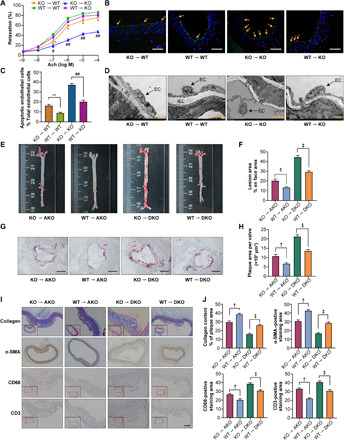
Fig. 3. BMT alleviated endothelial injury and atherosclerosis in mice.

As shown in fig. S4C, BMT was performed, and atherosclerosis was assessed after WD feeding for 12 weeks (10 mice in each group). (A) The aortic vasodilatation induced by Ach in KO mice (n = 10). (B) Representative images of TUNEL staining in sections of thoracic aortas. Scale bars, 200 μm. (C) The percentage of apoptotic endothelial cells (n = 5). (D) Representative electron microscopy images of endothelium in KO mice (n = 5). Scale bars, 50 μm. (E) Representative images of en face atherosclerotic lesion areas in AKO and DKO mice. (F) Quantitative analysis of (E) (n = 5). (G) Representative images of the cross-sectional area of the aortic root in AKO and DKO mice. Scale bars, 500 μm. (H) Quantitative analysis of (G) (n = 8). (I) Representative immunohistochemical staining images of VSMCs, collagen, macrophages, and T lymphocytes in aortic plaques. Scale bar, 100 μm. (J) Quantitative analysis of (I) (n = 5). The data are presented as the means ± SEM. *P < 0.05 versus WT → WT and **P < 0.01 versus WT → WT; #P < 0.05 versus WT → KO and ##P < 0.001 versus WT → KO; †P < 0.01 versus WT → AKO; ‡P < 0.001 versus WT → DKO.
