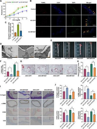
Fig. 4. The MYDGF overexpression of bone marrow in situ alleviated atherosclerosis.

In situ MYDGF overexpression in bone marrow was performed in KO, AKO, and DKO mice aged 4 to 6 weeks. Then, the mice were fed a WD for 12 weeks, and atherosclerosis was assessed at the end of the experiment (10 mice in each group). (A) The aortic vasodilatation induced by Ach in KO mice (n = 10). (B) Representative images of TUNEL staining in sections of thoracic aortas. Scale bars, 200 μm. (C) The percentage of apoptotic endothelial cells (n = 9). (D) Representative electron microscopy images of endothelium. Scale bars, 50 μm. (E) Representative images of en face atherosclerotic lesions. (F) Quantitative analysis of (E) (n = 5). (G) Representative images of the cross-sectional area of the aortic root. Scale bars, 500 μm. (H) Quantitative analysis of (G) (n = 9). (I) Representative immunohistochemical staining images of VSMCs, collagen, macrophages, and T lymphocytes in aortic plaques. Scale bar, 100 μm. (J) Quantitative analysis of (I) (n = 5). The data are shown as the means ± SEM. *P < 0.05 versus KO-GFP and **P < 0.001 versus KO-GFP; †P < 0.001 versus AKO-GFP; ‡P < 0.001 versus DKO-GFP.
