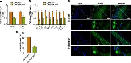
Fig. 5. The MYDGF overexpression of bone marrow in situ decreased the leukocytes homing within aortic plaques from DKO mice.

MYDGF overexpression of bone marrow in situ was performed in DKO mice aged 4 weeks, and leukocyte homing was analyzed in DKO-GFP or DKO-MYDGF mice that had been fed a WD for 12 weeks. (A) The mRNA expression of the macrophage markers F4/80 and CD68 in aortas. (B) The mRNA expression of the chemokines in aortas. (C and D) The homing of GFP leukocytes to atherosclerotic plaques 48 hours after intravenous injection into DKO-GFP and DKO-MYDGF mice that were fed a WD for 12 weeks. (C) Fluorescence micrograph of aortic root plaques. The dashed line indicates the plaque border. Inset, magnification of GFP leukocytes. Left, DAPI; middle, GFP; right, merge. Scale bars, 150 μm. (D) Quantification of GFP leukocytes per square millimeter of plaque (n = 5). The data represent the means ± SEM; *P < 0.001.
