
Fig. 5. V2R and OTR binding pockets: Binding of AVP versus retosiban.
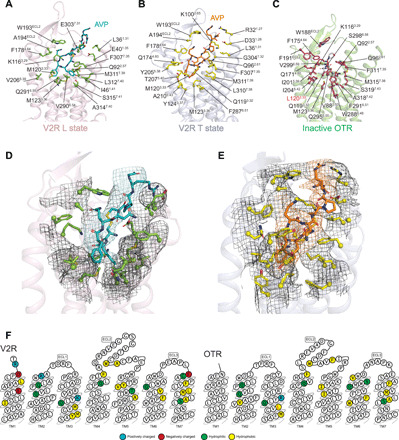
AVP binding poses are viewed from the side of the V2R helix bundle in L (A) or T (B) state and are compared with that of retosiban (in white sticks) in OTR (C). Receptor residues directly interacting with the ligands (at a maximum of 5 Å in distance) are indicated (Ballesteros-Weinstein numbering). In the OTR, L1203.33 (highlighted in red) is a mutation introduced in the sequence to increase thermostability and facilitate crystallogenesis (V120L). Densities of V2R residues in contact with AVP are shown in L (D) and T (E) states, respectively. (F) Residues of V2R and OTR involved in the binding of ligands are shown in receptor snake-like plot representations (https://gpcrdb.org).