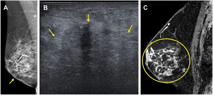
Figure 4.
70-year-old woman who presented with a self-detected mass in the right lower inner quadrant diagnosed on core-needle biopsy as invasive lobular carcinoma (ILC) (Nottingham grade 1, estrogen and progesterone receptor positive, HER2-negative) with associated lobular carcinoma in situ, which serves as an example of concordant estimation of the span of ILC on MRI with underestimation on conventional imaging. A: Diagnostic mediolateral oblique mammogram demonstrated skin thickening at the site of concern (arrow), but no definite mass. B: Targeted US demonstrated an ill-defined irregular-shaped mass with indistinct margins (arrows) that was difficult to measure but estimated to span 40 mm. C: MRI performed after biopsy demonstrated a total span of 92 mm of non-mass enhancement (circle) involving the lower inner and lower outer quadrants. Final pathologic size was 90 mm on mastectomy. Conventional imaging underestimated size based on the 5 mm, 25%, and T category of stage thresholds, while MRI was accurate using all three thresholds.

