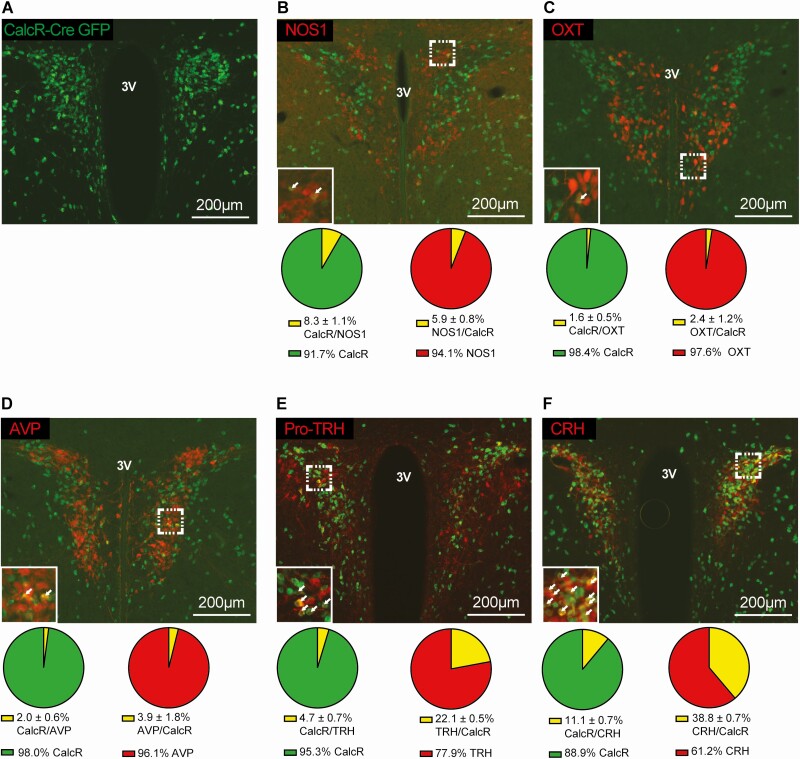
Figure 3.
Colocalization of CalcR PVH neurons with other PVH populations implicated in energy homeostasis. Representative immunofluorescent images taken of the PVH of CalcR2ACre+GFP mice. Mice were stained for Cre-dependent GFP expression (A-F) shown in green. Sections were also stained using primary antibodies for NOS1 (B), OXT (C), AVP (D), Pro-TRH (E), and CRH (F) shown in red. Percent expression of the overlap (yellow) relative to the individual cell populations (green/red) were quantified in corresponding pie charts below their image. Abbreviation: 3V, third ventricle.

