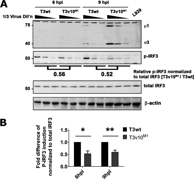
FIG 8.
T3v10M1 infection reduces levels of IRF-3 phosphorylation compared to that by T3wt. (A) L929 cells were exposed to T3wt or T3v10M1at a viral dose to infect 70% of the cells, which was confirmed by flow cytometry. Cell lysates were collected at 6 and 9 hpi. Total levels of IRF3 and phosphorylated (active form) IRF3 were determined by Western blotting. (B) Quantification of IRF3 activation. Levels of phosphorylated IRF3 (p-IRF3) were normalized to total IRF3. Three independent experiments were performed and graphed (mean ± SD; two-way ANOVA with Dunnett’s multiple-comparison test). *, P < 0.05; **, P < 0.01; ***, P < 0.001; ****, P < 0.0001.

