FIG 9.
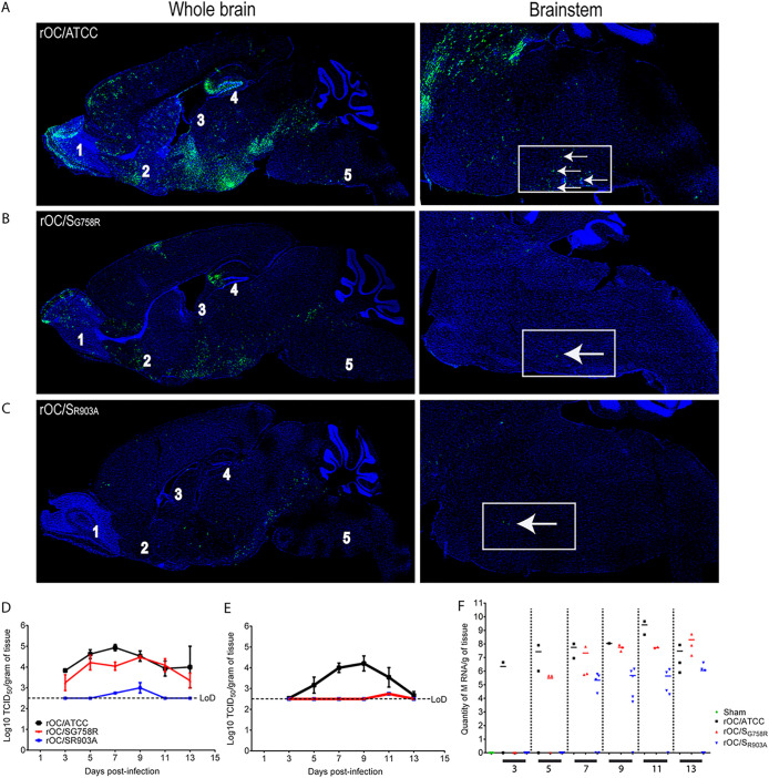
rOC/SR903A mutant virus spread in the CNS is abrogated compared to that of rOC/ATCC and rOC/SG758R. (A to C) Histological examination of virus spread within the brains of 22-day-old C57BL/6 mice infected with 101.5 TCID50/10 μl of rOC/ATCC (A), rOC/SG758R (B), or rOC/SR903A (C) by the i.c. route. Detection of viral antigens in whole brain (left) or in the brain stem (right) of infected mice at 7 dpi is shown. White arrows represent infected cells that were stained in green with MAb against the S viral glycoprotein. Blue staining is DAPI staining the nucleus (DNA). Magnifications are ×10 for left images and ×20 for right images. 1, olfactory bulb; 2, pyriform cortex; 3, lateral ventricle; 4, hippocampus; 5, brain stem. (D and E) Production of infectious viral particles was measured in brains (D) and spinal cords (E) every 2 days between 3 and 13 dpi. Results are the mean values (with standard deviations) from three independent experiments. (F) In spinal cord samples where no infectious virus was detected, RNA of each virus was quantified by qRT-PCR between 3 and 13 dpi after i.c. injection in C57BL/6 mice. Every symbol represents one single infected mouse.

