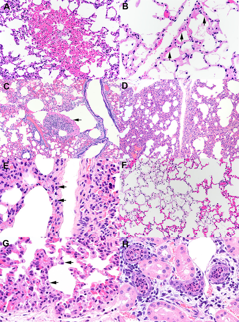
FIG 8.
Histological examination of lung from white-tailed deer fawns intranasally inoculated with SARS-CoV-2. (A) Note the well-demarcated focus of congestion. Alveolar septal capillaries are engorged with blood surrounded by normal appearing alveolar septa. HE. (B) Multiple alveolar septa are lined by bands of eosinophilic hyalinized proteinaceous material (arrows) consistent with hyaline membranes. Multiple alveoli contain flocculent to fibrillar eosinophilic material consistent with fibrin. HE. (C) Expanded alveolus contains a large collection of fibrin, inflammatory cells, and cell debris (arrow). HE. (D) Alveolar septa are expanded by an inflammatory infiltrate (interstitial pneumonia) composed primarily of lymphocytes (arrows) and macrophages (E). HE. (F) Within a field of congested alveolar septa are irregular regions characterized by hypocellular septa containing few erythrocytes. Septal stroma is fibrillar and lightly eosinophilic. Multiple alveoli within these regions contain flocculent strands of fibrin. HE. (G) There is type II pneumocyte hyperplasia and an increase in alveolar macrophages (arrows). HE. (H) Lumens of cortical tubules in the kidney are filled with necrotic cellular debris. Renal tubules are variably lined by attenuated epithelium, occasionally have hypereosinophilic cytoplasm and pyknotic nuclei (degeneration and necrosis), and overall exhibit increased cytoplasmic basophilia (regeneration). Tubules are separated by interstitial edema and a cellular infiltrate composed of lymphocytes, plasma cells, and fewer macrophages. HE. Tissue sections were examined on day 21 postinoculation.

