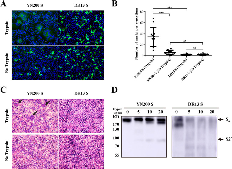
FIG 2.
The S protein induced syncytium formation and trypsin sensitivity. (A) Vero cells were transiently transfected with PCAGGS expressing YN200 S or DR13 S for 24 h. Cells were either left untreated or treated with trypsin for another 18 h and were subsequently monitored by immunofluorescence staining against the S protein (green). Nuclei were stained with DAPI (blue). (B) The numbers of nuclei per syncytium were determined and are displayed in the scatter plot. Asterisks indicate significant differences (**, P < 0.01; ***, P < 0.001); ns, not significant. (C) 293T cells were transfected with PCAGGS encoding the S protein. After 24 h, cells were removed from the plate and collected. Then the samples were lysed by ultrasonication before treatment for 1 h with 0, 5, 10, or 20 μg/ml of trypsin at 37°C. Cleaved S proteins were detected by Western blotting with a C-terminal His tag antibody. S0, full-length S protein; S2*, a cleavage product.

