FIG 5.
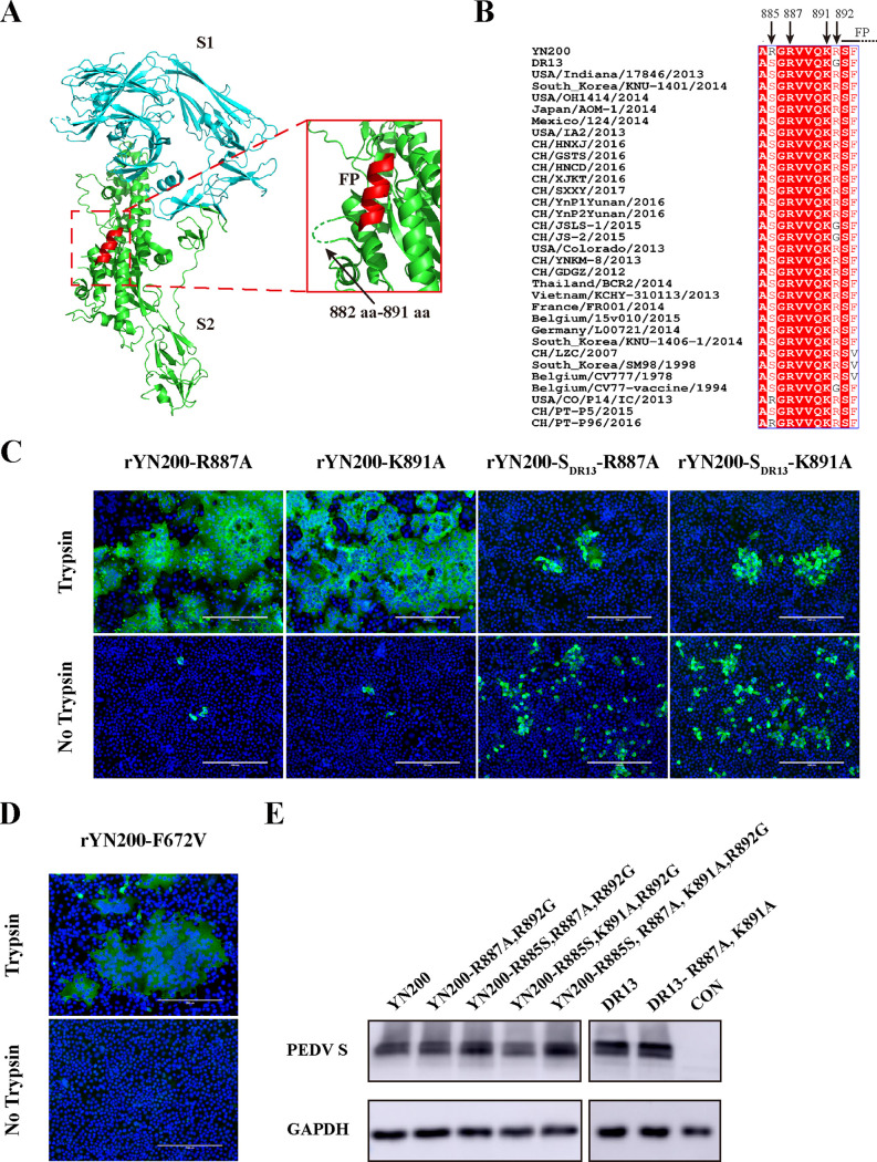
Structure and sequence analysis of the PEDV S protein. (A) Three-dimensional structure of the PEDV S protein monomer from the Protein Data Bank (PDB accession number 6U7K). S1, S2, and FP are color-coded cyan, green, and red, respectively. (Inset) Amino acids 882 to 891 are represented by a dashed green line. (B) Multisequence alignment of PEDV S sequences in the vicinity of the proposed S2′ position. Arrows indicate the amino acids of four critical sites. (C) Vero cells were infected with either rYN200-R887A, rYN200-K891A, rYN200-SDR13-R887A, or rYN200-SDR13-K891A with or without trypsin for 18 h (MOI, 0.01). Cells were examined by immunostaining against the nucleocapsid protein (green), and nuclei were stained with DAPI (blue). (D) rYN200-F672V was characterized by the same procedure as that used for panel C. (E) 293T cells were transfected with PCAGGS encoding the S protein. After 24 h, the S proteins were detected by Western blotting using the anti-S1 antibody or the anti-GAPDH antibody. CON, empty vector control.

