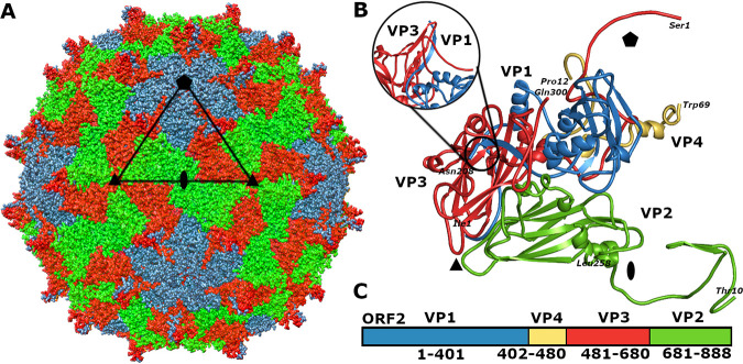
FIG 1.
Structure of KBV virion. (A) Molecular surface representation of KBV virion with subunits VP1 shown in blue, VP2 in green, and VP3 in red. The borders of one icosahedral asymmetric unit are outlined with a black triangle. The positions of selected symmetry axes are indicated with a pentagon for fivefold, triangle for threefold, and oval for twofold. Bar, 10 nm. (B) Cartoon representation of icosahedral asymmetric unit of KBV. VP1 subunit is shown in blue, VP2 in green, VP3 in red, and VP4 in yellow. The positions of icosahedral symmetry axes are indicated as in panel A. The inset shows a side view of a spike formed by a CD loop of VP3 and C-terminal β-strand VP1. (C) Organization of open reading frame 2 of KBV. Capsid proteins are colored as in panel B.

