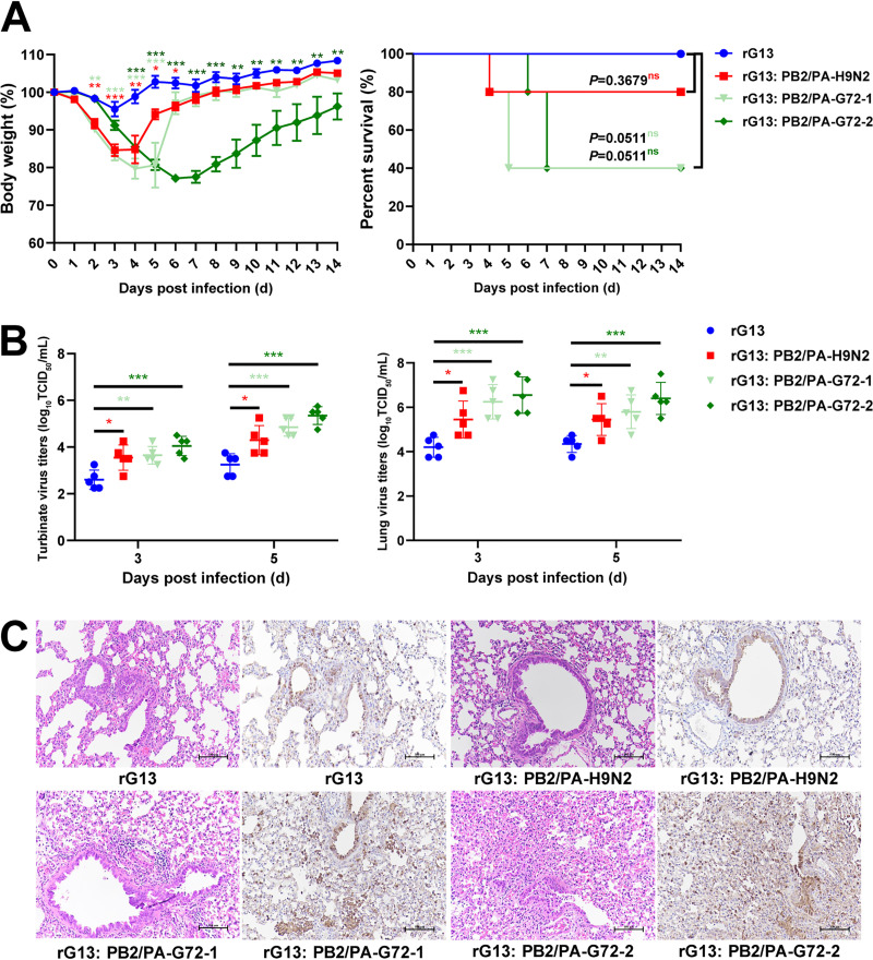
FIG 7.
Contribution of PB2 and PA genes of H9N2 virus origin to infectivity of H7N9 virus in mice. (A) Weight loss and percent survival of mice (n = 5) inoculated with 106 TCID50 of each virus. Mice that lost >25% of their baseline weight were euthanized. (B) Virus production from nasal turbinates and lungs. Five mice from each group were euthanized at 3 and 5 dpi to determine viral titers in nasal turbinates and lung tissues. Statistical significance was based on one-way ANOVA (*, P < 0.05; **, P < 0.01; ***, P < 0.001). (C) Representative histopathological changes in lung sections at 3 dpi by hematoxylin and eosin (H&E) staining (left) and immunodetection of influenza viral NP antigen (right). Mice infected with rG13:PB2/PA-H9N2, rG13:PB2/PA-H7N9-1, and rG13:PB2/PA-H7N9-2 viruses presented with more severe histopathology and a greater abundance of viral antigen-positive cells in the lung fields.

