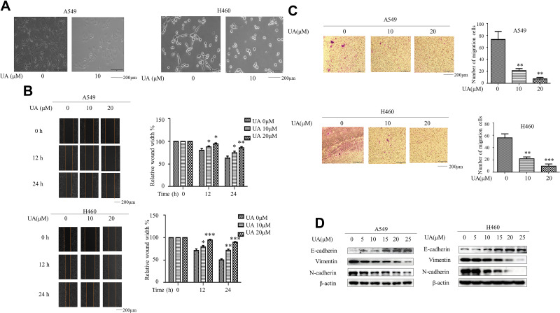
Figure 1.
Urolithin A inhibits EMT in lung cancer cells. (A) A549 and H460 cells were exposed to 10 μM urolithin A for 24 h. Cell morphology was assessed by phase-contrast microscopy (magnification 100×), scale bars 200 μm. (B) Wound Healing Assay reveals a dose-dependent (urolithin A 0, 10, 20 μM) (upper panel) and time-dependent (0, 12, 24 h) (lower panel) decrease in cell migration, scale bars represent 200 μm. The quantification was present in right panel. (*P < 0.05, **P < 0.01, ***P < 0.001 for difference from untreated control by ANOVA with Dunnett’s correction for multiple comparisons). (C) A549 and H460 were exposed to different concentrations of urolithin A (0, 10, 20 μM) treatment for 24 h. The cell invasiveness was assessed by Transwell chamber, scale bars indicate 200 μm. (**P < 0.01, ***P < 0.001 for difference from untreated control by ANOVA with Dunnett’s correction for multiple comparisons). (D) A549 and H460 cells were treated with different concentrations of urolithin A (0, 5, 10, 15, 20, 25 μM). Western blot examined epithelial marker E-cadherin, mesenchymal markers N-cadherin, Vimentin.

