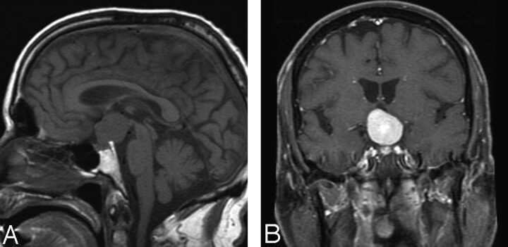
Fig 1.
A, Sagittal MR image shows a rounded mass in the suprasellar region, extending to the third ventricle. B, Coronal enhanced scan shows intense enhancement of the mass lesion, which appears to originate in the suprasellar region with fairly well-defined margins. The pituitary stalk, however, is not seen as a structure separate from the mass.

