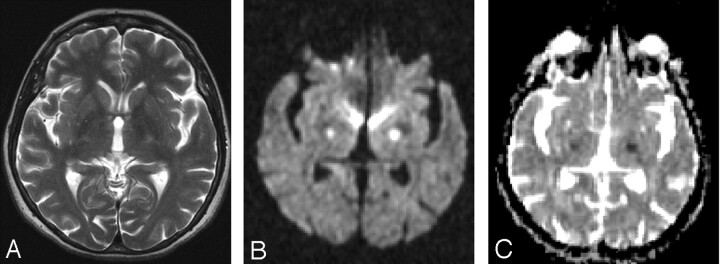
Fig 2.
MR imaging was performed at 12:00 am on August 13; glucose level was 32 mg/dL at 11:00 am. MR imaging was performed when the patient had hemiparesis. T2-weighted MR imaging (TR/TE, 5810/116 ms; number of excitations, 2) (A) shows suspected hyperintensities within the bilateral internal capsule. Diffusion-weighted imaging (B) (b = 1000 s/mm2; TE, 110; gradient strength, 24 mT/m) shows the presence of hyperintense lesions within the bilateral internal capsule. ADC values (C) calculated this time for the left internal capsule are 0.43 min/0.47 max/0.44 avg 10–3 mm2/s and, for the right internal capsule, are 0.50 min/0.55 max/0.52 avg 10–3 mm2/s.

