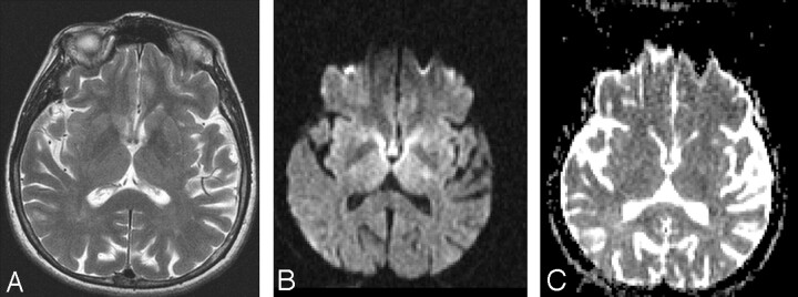
Fig 3.
MR imaging was performed at 7:00 pm on August 13; glucose level was 80 mg/dL at 6:00 pm. The patient’s hemiparesis improved immediately after the glucose infusion, and she recovered completely within hours without neurologic deficit. T2-weighted MR image (TR/TE, 4000/116 ms; number of excitations, 2) (A) shows no signal intensity changes. Diffusion-weighted MR image (b = 1000 s/mm2; TE, 113; gradient strength, 24 mT/m) (B) after recovery shows prominent regression of hyperintense lesions within the bilateral internal capsule, and ADC values (C) for the left internal capsule are 0.61 min0.73 max/0.65 avg 10–3 mm2/s and, for the right internal capsule, are 0.64 min/0.70 max/0.60 avg 10–3 mm2/s.

