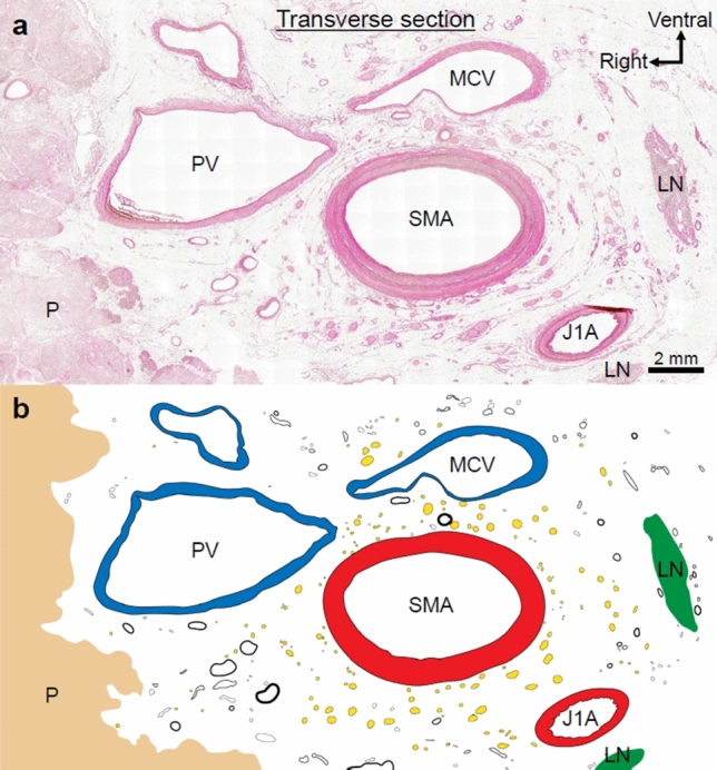
Fig. 3.

a Transverse histological section of the plate-like structure (hematoxylin and eosin stain. b Nerves, vessels (arteries, veins, and lymph ducts), collagen fibers, and adipose tissue existed in the region between the uncinate process of the pancreas and the SMA (nerves are indicated by yellow dots; vessels are indicated by circles). Collagen fibers encircled the SMA concentrically and wrapped around the nerves and small vessels in multiple layers to form the SMA plexus. LN lymph node, P pancreas, PV portal vein, MCV middle colic vein, SMA superior mesenteric artery, J1A first jejunal artery
