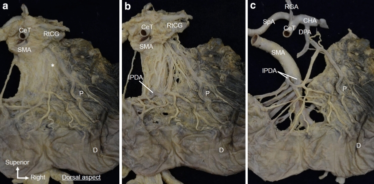
Fig. 6.
Detailed macroscopic analysis of the isolated en bloc specimen observed from the dorsal side. a Dorsal side of the same specimen shown in Fig. 5. The plate-like structure (*) is continuously connected to the RtCG, CeT, SMA, and pancreatic head. b After removing the loose connective tissue from the plate-like structure, the fibrous bundles of the plate-like structure were observed to spread radially from the RtCG and the roots of the CeT/SMA toward the pancreatic head. Some fibrous bundles were observed running along the IPDA. c After removing other arteries (b), arterioles branching from the dorsal pancreatic artery were observed within the plate-like structure. CeT celiac trunk, CHA common hepatic artery, D duodenum, DPA dorsal pancreatic artery, IPDA inferior pancreaticoduodenal artery, P pancreas, RtCG right celiac ganglion, SMA superior mesenteric artery, SpA splenic artery

