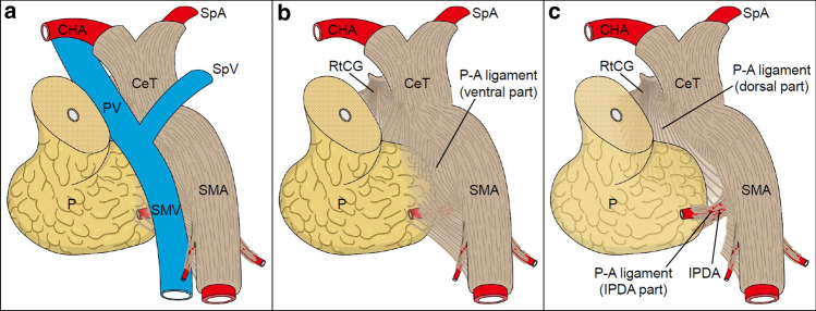
Fig. 7.
Schematic diagram of the P–A ligament. a Ventral view (slightly inferior and right) of the pancreatic head and surrounding structures. The pancreatic head is pulled slightly to the right, as it appears during surgery. b Region posterior to the PV/SMV. The P–A ligament is continuously connected to the RtCG, CeT/SMA, and pancreatic head and is composed of macroscopically visible fibrous bundles and loose connective tissue. In the ventral part of the P–A ligament, the uncinate process of the pancreas is attached to the SMA plexus, and the fibrous bundles mainly run along the SMA. c View of the region deeper (dorsal) than that in (b). In the dorsal part of the P–A ligament, the fibrous bundles spread radially from the RtCG and CeT toward the pancreatic head and the dorsal side of the SMA. Some fibrous bundles entered the pancreatic head along with the IPDA. P–A pancreas–major arteries, CeT celiac trunk, CHA common hepatic artery, IPDA inferior pancreaticoduodenal artery, P pancreas, PV portal vein, RtCG right celiac ganglion, SMA superior mesenteric artery, SMV superior mesenteric artery, SpA splenic artery, SpV splenic vein

