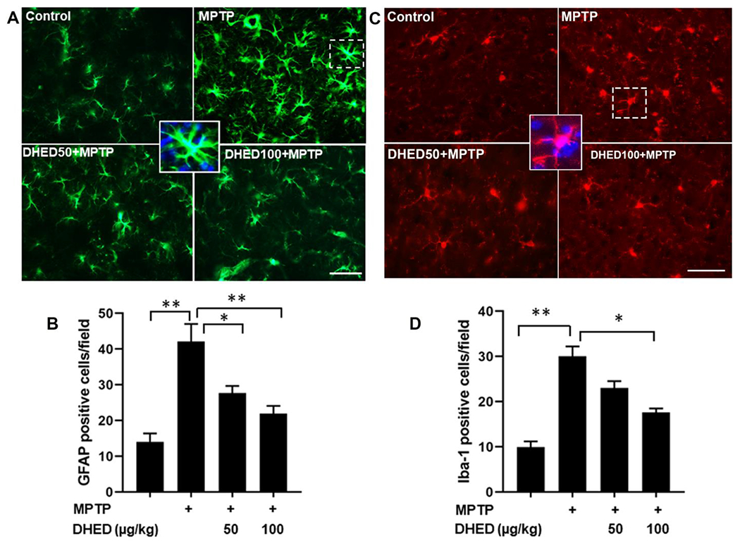
Fig. 3. DHED treatments inhibits glia cells activation in MPTP-treated mice.

The expression of microglial (Iba1) and astrocytic (GFAP) markers was determined by immunofluorescence in the brains of mice of each group. The upper panel shows representative fluorescent images of GFAP (A) and Iba1 (C) in striatum. The lower panel shows quantitative analysis of GFAP (B) and Iba1 (D) in brain. The profound expression of Iba-1 and GFAP (green color) were observed in MPTP group as compared to control group, while the MPTP group treated with DHED has shown a moderate staining of Iba-1 and GFAP. However, the control group has shown reduced staining. Scale bar, 50 μm. The values are expressed as mean ± SEM *P < 0.05; **P < 0.01 (N=3/group).
