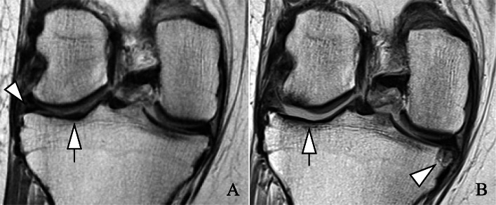
Fig. 2.
Severe progression of right knee degenerative changes over 4 years in an opioid user. Images were reviewed on picture archiving and communication system (PACS) workstations. Coronal views from intermediate-weighted sequences. a Baseline exam. Beginning extrusion and intra-substance lesion of the lateral meniscal body (arrowhead) and cartilage signal abnormality at the lateral tibial plateau (arrow). b Deterioration and progressive extrusion of the lateral meniscal body. Full-thickness cartilage loss at the medial femoral condyle (> 1 cm) and medial tibial plateau (> 1 cm) (arrow). Of note, new partial thickness cartilage loss at the medial femoral condyle and new, large subchondral cyst at the medial tibial plateau (arrowhead)

