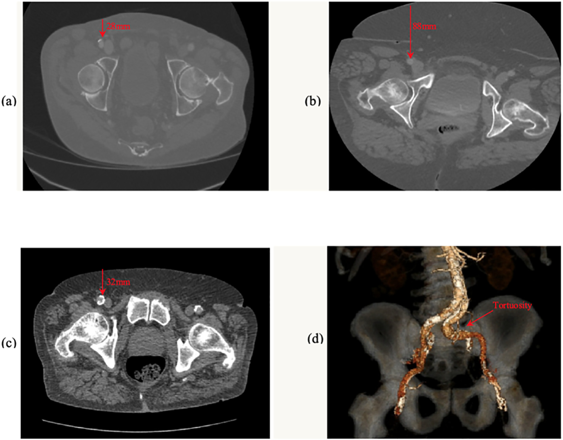
Fig. 2.

Computed tomography images of (a) non-contrast axial image of a shallow (28 mm depth) right CFA with anterior calcification, (b) a deep (88 mm) right CFA without calcification, (c) a relatively shallow right CFA with near complete circumferential calcification and (d) a 3-dimensional reconstructed view demonstrating marked tortuosity of the left common iliac artery. The red arrows in the figures indicate the depth (mm) and location of the corresponding CFA.
