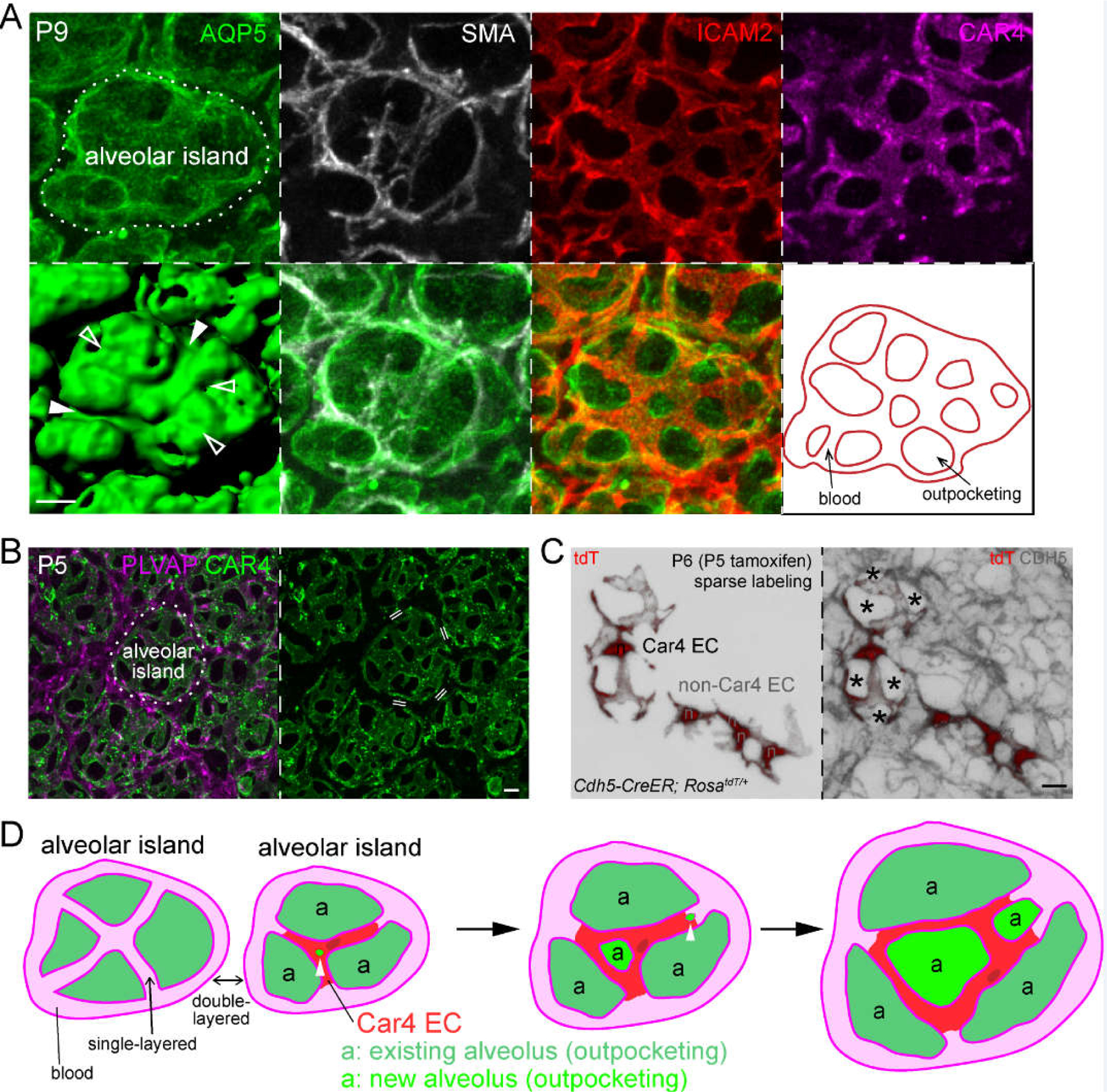
Figure 3: Car4 endothelial cells (ECs) cover alveolar islands and a model that links alveolar outpocketing to intussusceptive angiogenesis with implications for single-versus double-layered capillaries.

(A) En face view of confocal images of an immunostained alveolar region (AQP5) showing grooves with (filled arrowhead) or without (open arrowhead) alveolar myofibroblasts (marked by SMA), but invariably associated with vessels (ICAM2; also traced for the indicated alveolar island in the last panel). Car4 endothelial cells (CAR4) are specifically located over the alveolar island.
(B) En face view of confocal images of an immunostained alveolar region showing Car4 ECs specifically cover alveolar islands, but do not surround them as Plvap ECs do. Double line: double-layered capillaries are between alveolar islands, whereas vessels on alveolar islands are single-layered.
(C) En face view of confocal images of an immunostained alveolar region with individual endothelial cells (n, nucleus) sparsely, genetically labeled with a RosatdT reporter. The Car4 EC, as identified by costaining in the original publication,43 has cellular extensions contributing to 5–10 vessel segments, which are marked by Cadherin 5 (CDH5). Asterisks: alveolar outpocketings. Non-Car4 ECs have limited projections. (A-C) are adapted from43.
(D) A model depicting 2 alveolar islands with associated vessels and alveoli (outpocketings). The alveolar island on the right is shown to form 2 new alveoli, possibly coinciding with intussusception (arrowhead). Depending on whether intussusception occurs in the middle or at the cell junction of a Car4 EC (darker patch: nucleus), the resulting Car4 EC forms an auto-cellular loop or cellular extensions, respectively. Such a model of alveologenesis does not involve bending capillaries into a hairpin conformation so that single-layered capillaries on alveolar islands remain single-layered but increase in number as a result of intussusception, which dilutes the double-layered capillaries between alveolar islands over time.
Scale: 10 um.
