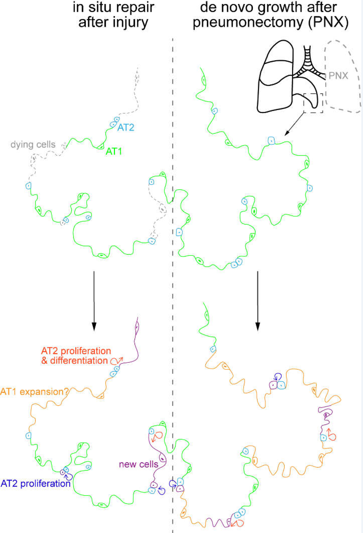
Figure 4: In situ repair versus de novo growth as distinct modes of lung regeneration.

Two extreme modes of lung regeneration. Left: epithelial cell loss without affecting the extracellular matrix can be replaced in situ via AT2 cell proliferation and their differentiation into AT1 cells, as well as possible AT1 cell expansion. Not depicted is the contribution of airway-derived cells, such as p63-expressing atypical basal cells forming dysplastic clusters. Right: In pneumonectomy, where the left lobe is removed, the uninjured lobe grows to compensate for the loss in gas exchange capacity. This growth in tissue volume, surface area, and cell number occurs de novo in the available empty space, possibly due to the reactivation of developmental programs. Cellular changes are color-coded as in the left schematic. Chemical or pathogen-induced lung injury likely activates both modes of lung regeneration.
