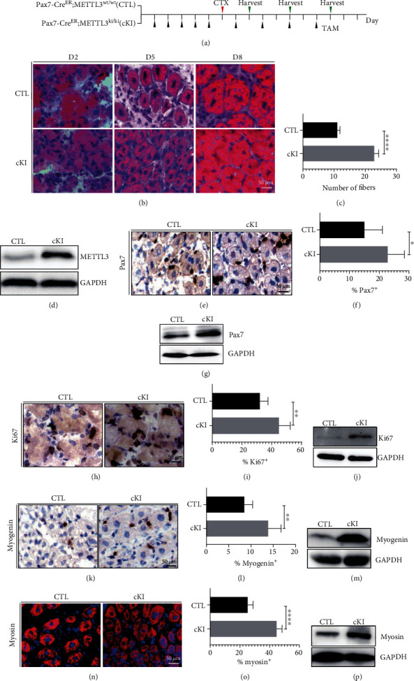
Figure 3.

METTL3 conditional knockin promotes muscle regeneration in vivo. (a) Schematic outline of CTX injection in TAM-treated control and METTL3 cKI littermates at the age of 8–9 weeks. (b) H&E staining of representative tibialis muscle sections from METTL3 cKI, control mice treated 2, 5, and 8 days after CTX injection. (c) Quantified of muscle fibers. (d) The upregulation effect of METTL3 was verified at protein levels by Western blot. (e, h, k) Immunohistochemical and (n) immunofluorescence staining of TA muscle from METTL3 cKI and control detected by Pax7, Ki67, myogenin, and myosin antibodies. (f, i, l, o) Quantifications of Pax7+, Ki67+, myogenin+, and myosin+ cells were shown on the right. (g, j, m, p) Western blot detected the protein expression levels of Pax7+, Ki67+, myogenin, and myosin. Data represent mean ± SEM. N = 5. ∗∗∗∗p < 0.0001, ∗∗p < 0.01, ∗p < 0.05. CTX: cardiotoxin; TAM: tamoxifen; TA: tibialis anterior; CTL: control; cKI: METTL3 conditional knockin.
