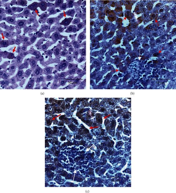
Figure 3.

Microphotographs of intracellular Nrf2 expression in mice's liver 30 days after BCG infection (×400). (a) Representative immunohistochemical staining for Nrf2 in livers from control mice. Arrows indicate Nrf2 intracellular localization (cytoplasmic but not nuclear). (b) Representative immunohistochemical staining for Nrf2 in livers from BCG-infected mice. Arrows indicate Nrf2 intracellular localization (cytoplasmic dominant) in hepatocytes (red arrows); granuloma cells are not stained (white arrows). (c) Representative immunohistochemical staining for Nrf2 in livers from BCG-infected mice that received TS-13 in drinking water. Arrows indicate Nrf2 intracellular localization (cytoplasmic and nuclear) in hepatocytes (red) and in granuloma cells (white).
