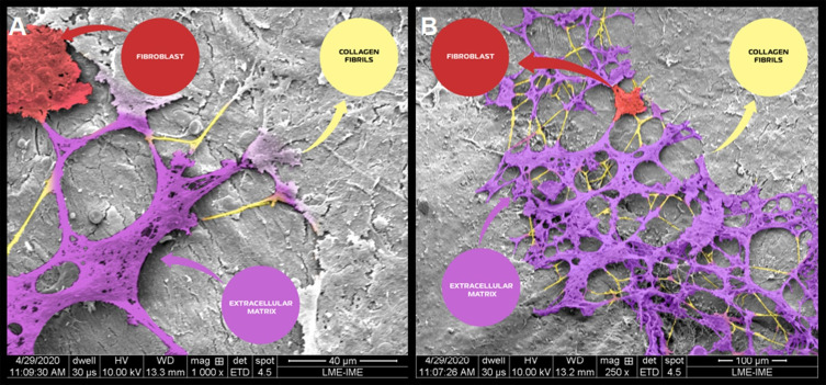
Figure 13.
Scanning electron micrographs of bone defects filled with nano-hydroxyapatite/ß tricalcium phosphate showing the surface of the bone matrix. The highlighting images are suggestive of collagen fibrils (yellow), collagen fibrils associated with other types of proteins (magenta) and possibly starting the process of the extracellular matrix formed by an aggregate of glycoproteins and bundles of collagen fibers; and a fibroblast (red). (A) scale bar = 40 µm, x1000; (B) scale bar 100 µm, X 250 magnification.

