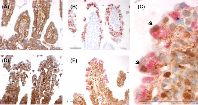
Figure 2.
NF-κB staining and C. suis staining in sections of the jejunum. The jejunum of an uninfected animal (A) or an animal infected with C. suis(B–E) at 2 days post-inoculation (dpi) is shown. Panels A and D show NF-κB single staining (brown), B shows C. suis single staining (red), C and E show NF-κB (brown) staining and C. suis (red) double staining. Panel C is a detail of Panel E. Scale bar 50 µm. Arrowheads indicate C. suis-infected cells with labeled nuclear NF-κB and asterisk indicates C. suis-infected cell with no labeled nuclear NF-κB.

