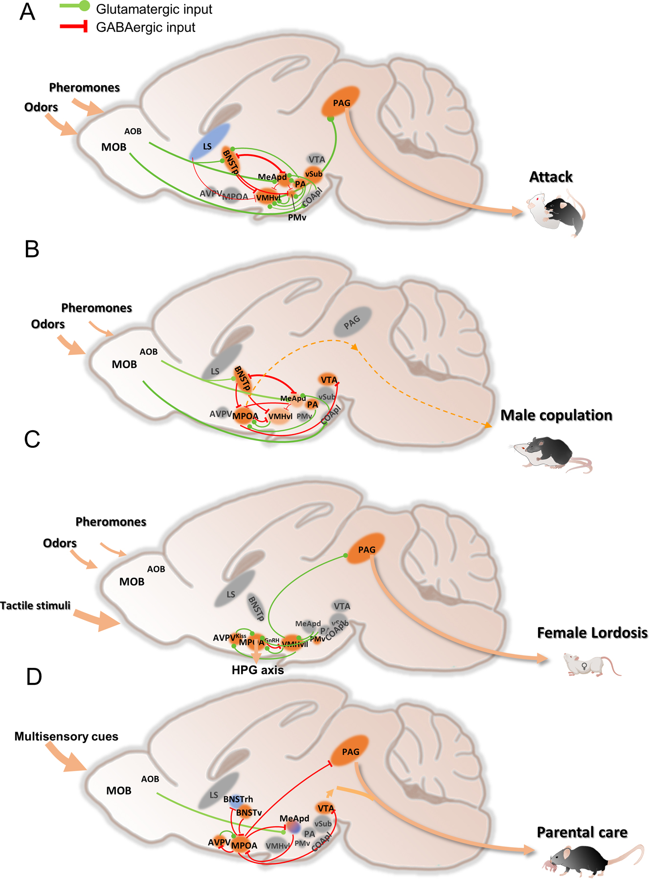
Figure 3. Schematics showing neural circuits of social behaviors.

(A) aggression circuit;
(B) male sexual behavior circuit;
(C) female sexual behavior circuit;
(D) parental care circuit.
Solid lines denote known pathways that are involved in mediating the behaviors. Dashed lines denote potential pathways to be further explored. Blue colored regions suppress the behavior while orange colored regions promote the behaviors. Light orange marks regions that play minor roles in the behavior. Gray colored regions are not essential for the behaviors or not yet studied. Arrow size indicates importance of the sensory input. Line width indicates connection strength. Not all the known connections are shown. AOB: accessory olfactory bulb; AVPV: anteroventral periventricular nucleus; BNSTp: posterior part of the bed nucleus of stria terminalis; BNSTrh: rhomboid nucleus of the bed nucleus of stria terminalis; BNSTv: ventral part of the bed nucleus of stria terminalis; COApl: posterolateral cortical amygdala; LS: lateral septum; MeA: medial amygdala; MPOA: medial preoptic area; PA: posterior amygdala; PAG: periaqueductal gray; PMv: ventral premammillary nucleus; VMHvl: ventrolateral part of the ventromedial hypothalamus; VMHvll: lateral subdivision of the VMHvl; vSUB: ventral subiculum; VTA: ventral tegmental area.
