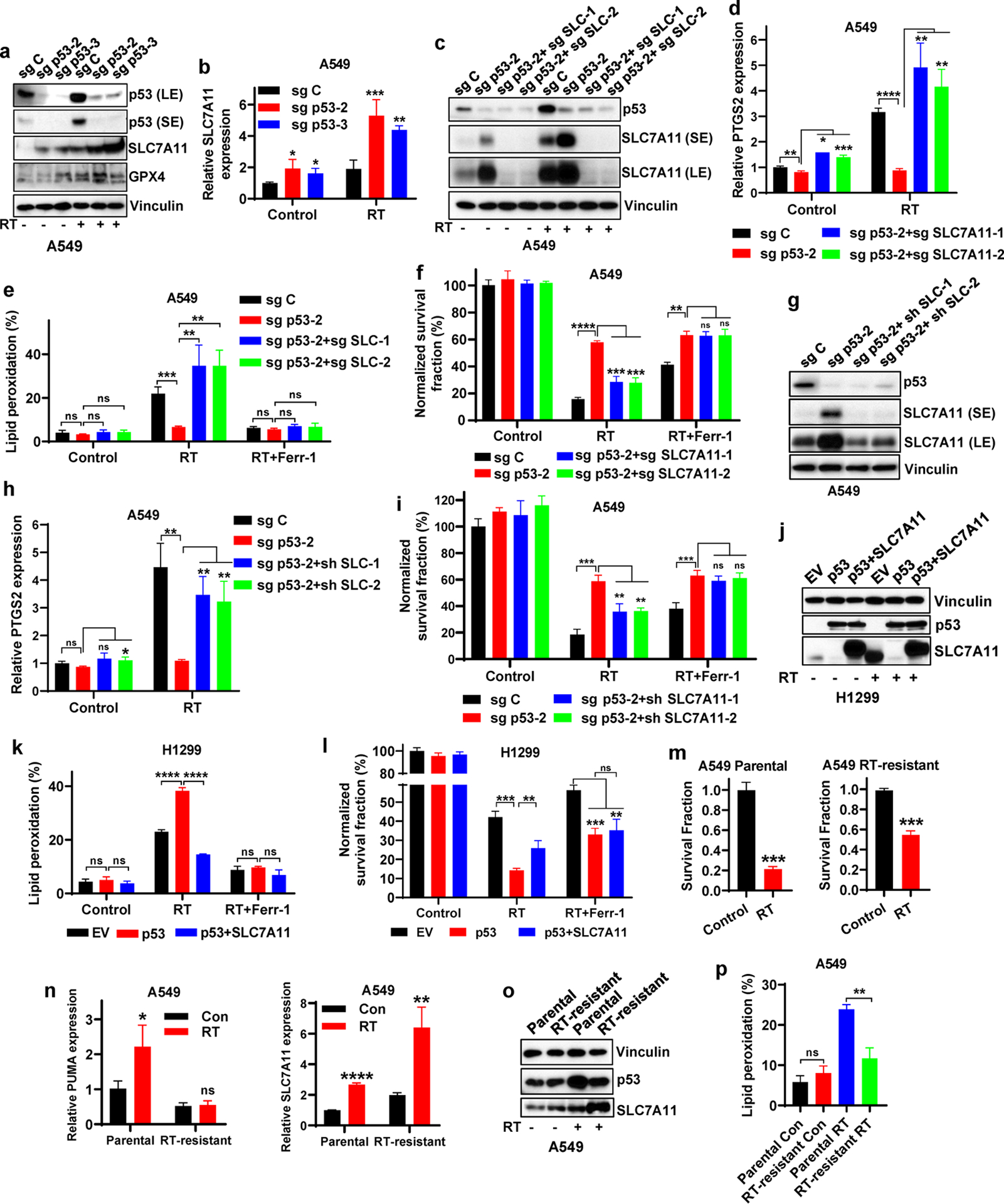
Figure 2. p53 promotes RT-induced ferroptosis partly via antagonizing SLC7A11 induction.

a Western blotting indicating p53, SLC7A11 and GPX4 levels in sg C, sg p53-2, and sg p53-3 A549 cells without X-ray irradiation or at 12 hours after 6 Gy X-ray irradiation.
b mRNA levels of SLC7A11 were analyzed by qPCR in sg C, sg p53-2, and sg p53-3 A549 cells without X-ray irradiation or at 12 hours after 6 Gy X-ray irradiation.
c Western blotting indicating p53 and SLC7A11 levels in sg C, sg p53-2, sg p53-2+sg SLC7A11-1 and sg p53-2+sg SLC7A11-2 A549 cells without X-ray irradiation or at 12 hours after 6 Gy X-ray irradiation.
d PTGS2 mRNA levels in sg C, sg p53-2, sg p53-2+sg SLC7A11-1, and sg p53-2+sg SLC7A11-2 A549 cells without X-ray irradiation or at 12 hours after 6 Gy X-ray irradiation.
e Lipid peroxidation analysis in sg C, sg p53-2, sg p53-2+sg SLC7A11-1, and sg p53-2+sg SLC7A11-2 A549 cells without X-ray irradiation or at 12 hours after exposure to 6 Gy X-ray irradiation following pretreatment with 5 μM ferrostatin-1 or DMSO for 24 h.
f Clonogenic survival analysis of sg C, sg p53-2, sg p53-2+sg SLC7A11-1 and sg p53-2+sg SLC7A11-2 A549 cells exposed to 6 Gy X-ray irradiation following pretreatment with 5 μM ferrostatin-1 or DMSO for 24 h.
g Western blotting indicating p53 and SLC7A11 levels in sg C, sg p53-2, sg p53-2+sh SLC7A11-1 and sg p53-2+sh SLC7A11-2 A549 cells.
h PTGS2 mRNA levels in sg C, sg p53-2, sg p53-2+sh SLC7A11-1, and sg p53-2+sh SLC7A11-2 A549 cells without X-ray irradiation or at 12 hours after 6 Gy X-ray irradiation.
i Clonogenic survival analysis of sg C, sg p53-2, sg p53-2+sh SLC7A11-1 and sg p53-2+sh SLC7A11-2 A549 cells exposed to 6 Gy X-ray irradiation following pretreatment with 5 μM ferrostatin-1 or DMSO for 24 h.
j Western blotting indicating p53 and SLC7A11 levels in H1299 cell line with stable expression of EV, wild-type p53 and wild-type p53 + SLC7A11.
k Lipid peroxidation analysis in EV-, wild-type p53-, and wild-type p53 + SLC7A11-expressing H1299 cells without X-ray irradiation or at 12 hours after exposure to 6 Gy of X-ray irradiation following pretreatment with 5 μM ferrostatin-1 or DMSO for 24 h.
l Clonogenic survival analysis of EV-, wild-type p53-, and wild-type p53 + SLC7A11-expressing H1299 cells exposed to 6 Gy of X-ray irradiation following pretreatment with 5 μM ferrostatin-1 or DMSO for 24 h.
m Clonogenic survival analysis of parental and RT-resistant A549 cells following 6 Gy of X-ray irradiation.
n mRNA levels of PUMA and SLC7A11 were analyzed by qPCR in parental and radioresistant A549 cells with or without 6 Gy X-ray irradiation.
o Western blotting indicating p53 and SLC7A11 levels in parental and radioresistant A549 cells.
p Lipid peroxidation analysis in parental and radioresistant A549 cells at 24 hours after 6 Gy X-ray irradiation.
The percentage values in the panel e, k and p refer to the percentages of cells with lipid peroxidation measured by BODIPY™ 581/591 C11 staining followed by FACS analysis. Error bars are mean ± SD from three independent repeats. P values calculated by 2-tailed unpaired Student’s t-test.
