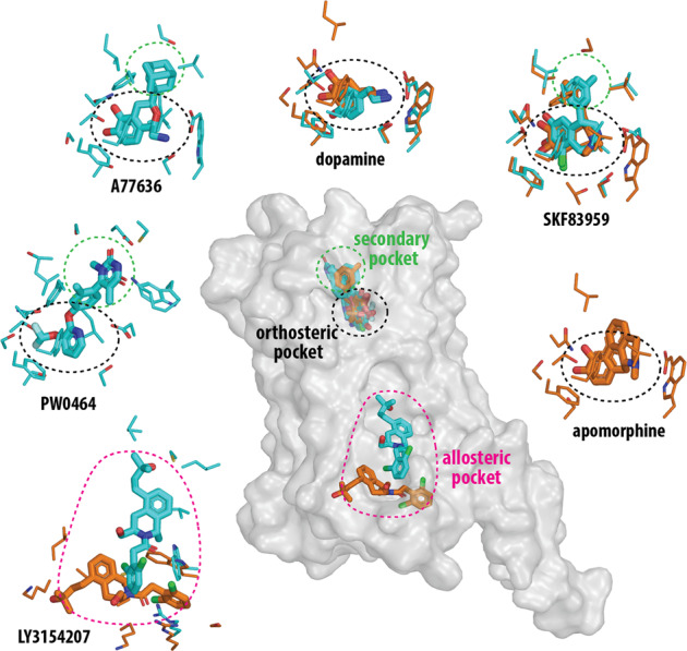
Fig. 1.

The DRD1 structures reveal orthosteric, secondary, and allosteric binding pockets. The relative locations of these pockets are indicated by colored ellipses in the central panel in which the active conformation of the DRD1 is shown in a transparent surface representation. The binding poses of selected ligands and their corresponding interacting residues (within 3.8 Å of the ligand) are shown in surrounding panels as labeled. The binding pockets are indicated by the ellipses in the same colors as the central panel. Those from Xiao, et al.1 are colored in cyan, those from Zhuang, et al.2,3 are colored in orange. In particular, three pairs of structures bound with dopamine, SKF83959, or LY3154207 from both groups are superimposed to demonstrate their divergences
