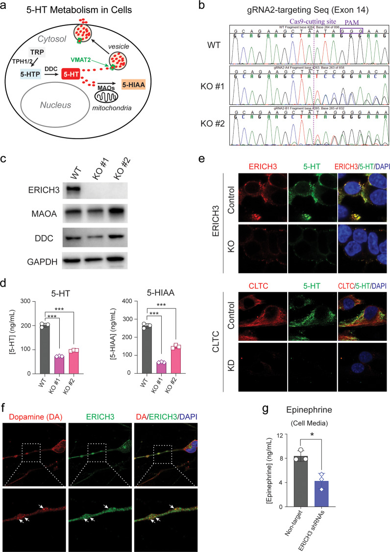
Fig. 4. ERICH3 and monoamine neurotransmitters.
a 5-HT is synthesized from tryptophan (TRP). Specifically, TRP is metabolized by tryptophan hydroxylases (TPH1 or TPH2) to generate 5-hydroxytryptophan (5-HTP) which can be further metabolized by aromatic-L-amino-acid decarboxylase (also known as DOPA decarboxylase, DDC) to form 5-HT. 5-HT can then be transported into vesicles by vesicular monoamine transporters (VMATs) and is released from cells by exocytosis. Vesicle-free 5-HT can be metabolized by monoamine oxidases (MAOs), which are located in the outer mitochondrial membrane to generate 5-hydroxyindoleacetic acid (5-HIAA). b Sanger sequencing of the ERICH3 exon 14 region, which was targeted by ERICH3 guide RNA 2 (gRNA2) in WT SK-N-SH cells, and two single colonies edited by CRISPR/Cas9 (KO#1 and KO#2). The Cas9-cutting site is labeled by dashed line and the protospacer adjacent motif (PAM), “GGG,” is boxed. Compared to WT cells, DNA sequences on the right side of the Cas9-cutting site shifted in KO#1 and KO#2 cells, indicating that the CRISPR/Cas9 editing occurred at the “designed” site in KO#1 and KO#2 cells. c Western blots showing that ERICH3 protein was not detectable and that MAOA and DDC protein levels were not significantly changed in the KO#1 or KO#2 cells when compared with WT cells. d 5-HT (left) and 5-HIAA (right) concentrations were significantly decreased in the cell culture media of SK-N-SH KO#1 and KO#2 cells in which the ERICH3 gene had been edited by CRISPR/Cas9. Cells were incubated with 40 µM 5-HTP for 6 h since SK-N-SH cells express neither TPH1 nor TPH2. Data are mean ± s.d. (n = 3), with statistical significance determined by Dunnett’s test denoted as ***P < 0.001. e Immunofluorescent (IF) costaining of ERICH3 and 5-HT showing the colocalization of ERICH3 with 5-HT in control SK-N-SH cells. In ERICH3 KO cells, 5-HT was hardly to detect by IF under the same conditions used to study control cells (upper panel). KD of CLTC resulted in a similar effect on 5-HT as did KO of ERICH3 (lower panel). Magnification is ×150. Figure shown represents results from duplicate experiments. f Costaining of ERICH3 and dopamine (DA) in human iPSC-derived dopaminergic neurons (hDN) showing the colocalization of ERICH3 with DA in neurites. Magnification is ×60 for upper panel and 300× for lower panel. Figure shown represents results from duplicate experiments. g Epinephrine, an end product of the dopamine metabolizing pathway was decreased in cell culture media for hDN 24 h after transfection with lentivirus packaged with ERICH3 shRNAs. Neither DA nor norepinephrine could be detected after 24 h incubation. Data are mean ± s.d. (n = 3), with statistical significance determined by unpaired t-test denoted as *P < 0.05.

