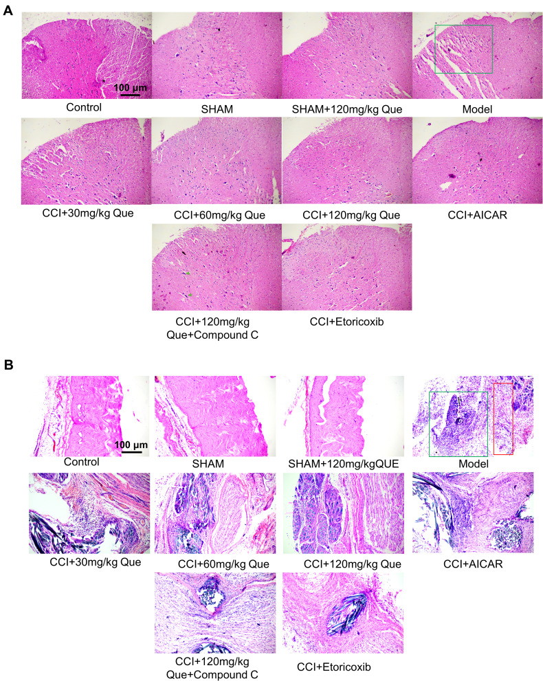
Figure 2.
(A) The HE staining of que on CCI-induced neuropathic pain on the 28th day after the ligation of the spinal cord. The green box is the dorsal horn area of the spinal cord, and the green arrow shows the large nissella body. (B) The HE staining of que on CCI-induced neuropathic pain on the 28th day after the ligation of sciatic nerve. The green box shows the presence of a large number of inflammatory cells, and the red box shows the location of the nerve.

