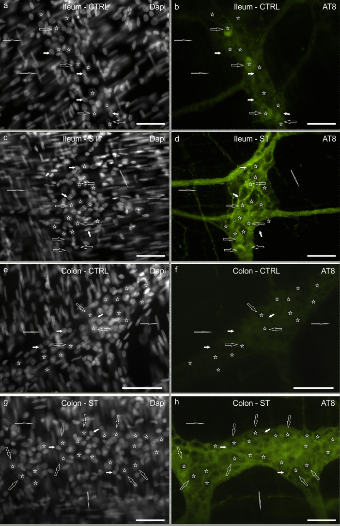
Fig. 1.
a–h Photomicrographs of whole-mount preparations of myenteric plexus (MP) of rat ileum (a–d) and colon (e–h) showing AT8 immunolabeling in control normothermic (CTRL) rats (a, b; e, f) and hypothermic (synthetic torpor, ST) rats (c, d; g, h). Open arrows indicate MP neurons that showed moderate-to-bright AT8 immunoreactivity in the ileum of CTRL rats (b) and faint AT8 immunoreactivity in the colon (f). In the ST rats, MP neurons (open arrows) showed bright AT8 immunoreactivity in both the intestinal segments (d, h). Stars indicate some of the neuronal nuclei stained with DAPI (a, c, e, g), which were recognizable for their shape, dimension and staining intensity. White arrows indicate the nuclei of some glial cells, which were smaller than the neuronal nuclei. Elongated open arrows indicate the nuclei of some smooth muscle cells. Bar: a–h = 50 µm

