Fig. 3.
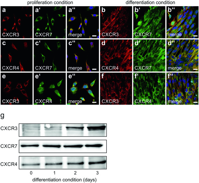
Expression and co-localization of chemokine receptors in C2C12 cells a–f C2C12 cells were maintained under either proliferation conditions a, c, e or differentiation conditions b, d, f for 3 days and double-labelled with antibodies against CXCR3 (bs-2209R), CXCR7, or CXCR4 as indicated. a″–f″ Merging of immunocytochemical stainings and DAPI. Virtually all non-differentiated C2C12 cells expressed CXCR3 a, e. Expression of CXCR3 persisted in forming myotubes b, f. In addition to CXCR3, again virtually all non-differentiated C2C12 cells as well as forming mytotubes co-expressed CXCR7 a″, b″ and CXCR4 e″, f″. Virtually all proliferating and differentiating cells further co-expressed CXCR4 and CXCR7 c″, d″. Scale bars, 10 µm. g Quantification of chemokine receptors expression in differentiating C2C12 cells. C2C12 cells were maintained for the indicated times with differentiation medium and analysed for CXCR3, CXCR7 and CXCR4 by Western blotting. Expression of the chemokine receptors increased with ongoing differentiation. Due to the lack of appropriate loading controls, care was taken to adjust samples to identical protein levels

