
Figure 4. Il6raMye−/− mice are more susceptible to liver fibrosis after 3-month HFD feeding.
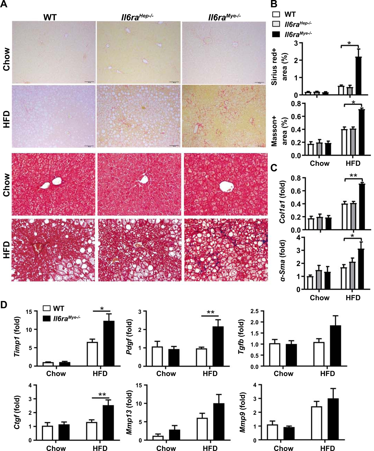
Il6raHep−/−mice, Il6raMye−/−mice, and WT mice were fed an HFD or chow for 3 months. (A) Representative images of Sirius red staining (upper two panels) and Masson staining (lower two panels) of liver tissue sections are shown. (B) Quantification of fibrotic area per field. (C, D) RT-qPCR analyses of hepatic fibrogenesis genes. Values represent means ± SEM (n = 11–14). *P< 0.05, **P< 0.01 in comparison with WT HFD groups.