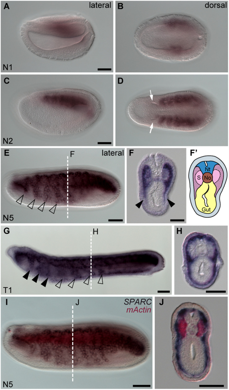
FIGURE 1.
Amphioxus SPARC is expressed in the lateral region (non-myotome part) of the somite. Images (A,C,E,G,I) were taken from the lateral view; the anterior is to the left and dorsal is to the top. Scale bars, 50 μm. Images (B,D) were taken from the dorsal view, where the anterior is to the left and the right side is on top. Scale bars in (A,C) apply to (B,D). Cross sections in panels (F,H,J) show the respective level marked with a white dashed line in panels (E,G,I). Scale bars, 25 μm. (E) SPARC expressing cells expand ventrally (hollow arrowhead). (F) Black arrowheads indicate the lateral part of the somite that expresses SPARC and extends ventrally. (F′) Schematic drawing of panel (F); area filled with dashed lines marks where SPARC is expressed. Nt, neural tube; S, somites; No, notochord. (G) SPARC-expressing cells move to the pharyngeal region (black arrowheads). (I) Double in situ hybridization of SPARC (dark purple) and mActin (red) of a late N5 stage embryo.

