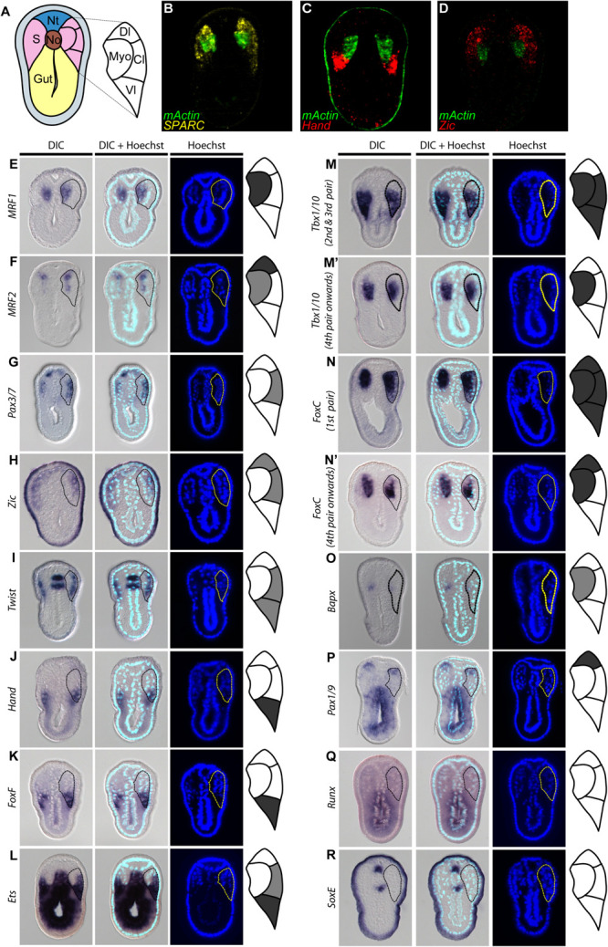
FIGURE 4.

Comparable compartments inside the amphioxus somite can be recognized by the expression of specific marker genes. (A) Schematic drawing of an N3 stage (six somite) embryo cross section. The somite region (pink) can be compartmentalized into four regions based on the expression of different marker genes. Myo, myotome; Dl, dorsolateral; Cl, Centrolateral; Vl, ventrolateral; Nt, neural tube; S, somite; No, Notochord. (B–D) Double fluorescent in situ hybridization of sectioned embryos. (E–R) Expression pattern of known somite marker genes in N3 stage embryos, taken from around 5th somite level. In the schematic diagrams, dark gray represents stronger expression while light gray represents weaker expression. The right somite is outlined with dashed lines. (Q,R) Expression patterns of known vertebrate sclerotome marker genes that are not expressed in the amphioxus somites. The right somite is outlined with dashed-lines.
